Breast Surgery

Breast Cancer

Breast cancer is the most common type of cancer affecting women globally, with 1 in 10 women developing it over their lifetimes. 81% of breast cancers occur in women over the age of 50. Exactly why some women get breast cancer while others don’t remain unknown. However, research suggests that breast cancer is caused by a combination of factors, some of which are beyond control.

Most breast cancers happen by chance. Despite this, approximately 5% of people diagnosed with breast cancer have an inherited genetic fault. Of the known breast cancer genes BRCA1, BRCA2, or TP53 are the most common. The three main risk factors for breast cancer are gender (being female), age (risk increases after age of 50), and significant family history (particularly when first-degree relatives are involved i.e., parent or sibling).

Other observed risk factors for breast cancer that have less significance, include a previous personal history of breast cancer, increased hormonal exposure such as early menarche (first period) or late menopause, and lifestyle (obesity, excessive alcohol intake, smoking, and diabetes).

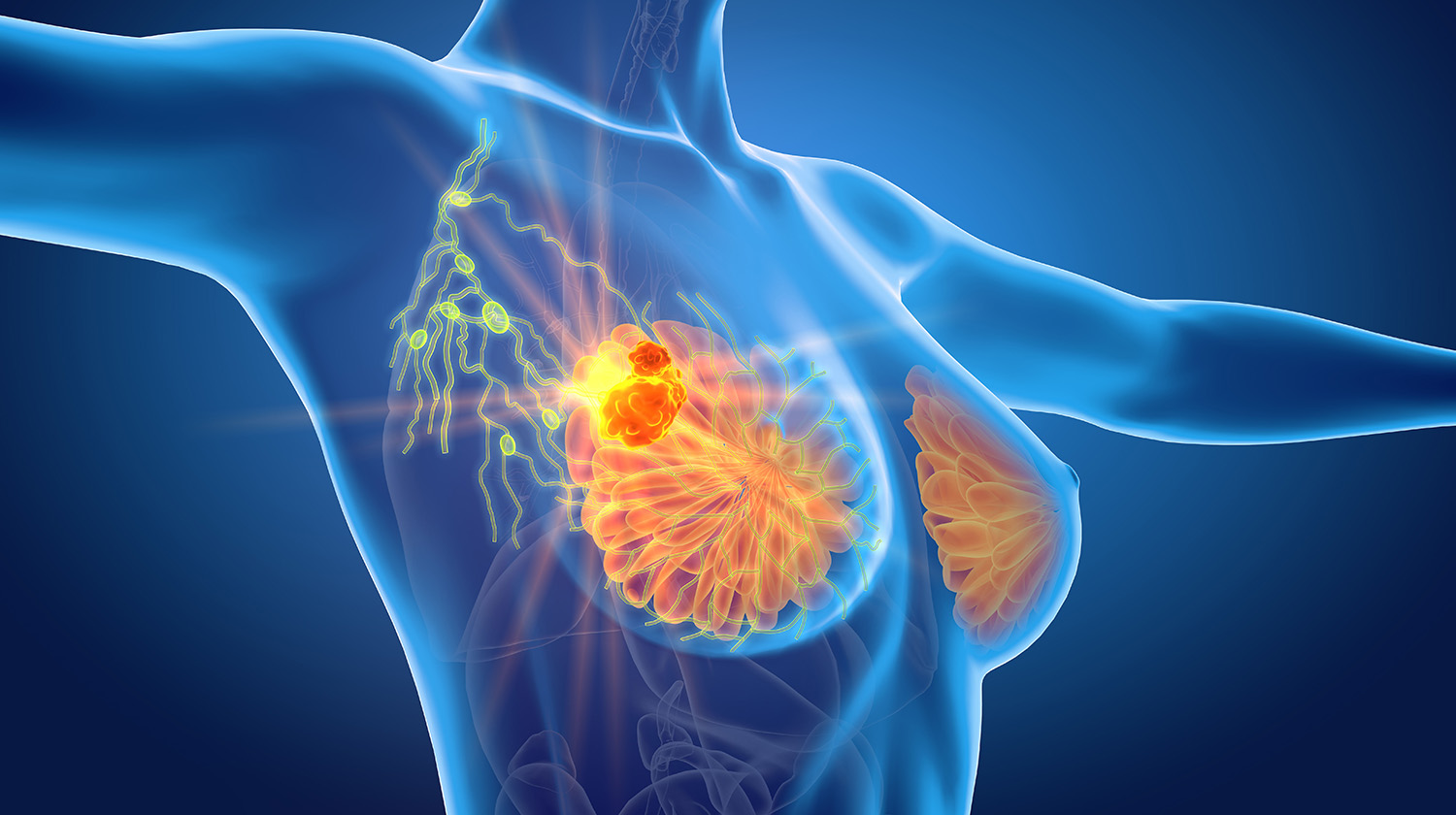
breast cancer

Breast Cancer

Diagnosis

The diagnosis of breast cancer is made using a combination of Clinical Examination (where a palpable lump is assessed by experienced medical personnel), 

a Radiological Examination (any combination of ultrasound, mammography, or MRI), and a Histological Assessment (where a biopsy is studied under a microscope).

Symptoms

  • In the early stages, breast cancer gives no symptoms and can only be detected by radiological assessment.
  • As the disease progresses, a lump may be felt - when the lump grows it may produce skin changes such as redness or dimpling.
  • As cancer grows, it will start to spread. The most common ‘first station’ is the lymph nodes of the armpit (axilla), and from there it can spread to distant locations (most commonly the liver, bones, and lungs).
  • A very large breast lump can ulcerate, bleed, or discharge fluid.
  • Nipple discharge can occasionally be an indicator of breast cancer.
  • Although a common complaint, breast pain is not a common presenting symptom of breast cancer.

Treatment

Surgical Treatment

  • Treatment for breast cancer depends on size, spread, previous history, and individual factors such as co-morbidities. Breast cancer treatment is often a combination of surgery, radiotherapy, chemotherapy, and hormonal therapy.
  • The treating specialist will form a plan by assessing each patient. Sometimes, this requires a discussion at a Multi-Disciplinary Team Meeting (MDT Meeting), where multiple specialists determine the most appropriate course of action.
  • Depending on the individual, other supporting specialists may be involved, including an occupational therapist, a physiotherapist, and a psychotherapist.
  • Most people start their treatment with surgery, then progress to other therapies dependant on the characteristics and status of the cancer.
Wide Excision

Wide Excision is the removal of cancer with a safety margin (where a small amount of tissue is taken around the tumour to ensure none is remaining in the body) and is followed by radiotherapy to eliminate any remaining cells. 
Patients who are good candidates for this surgery are those where the cancer was discovered in its early stages and is still small, and where the surgery can leave good cosmetic results.

Mastectomy

Mastectomy is the removal of the whole breast. This approach is usually used if the cancer is too big for a wide excision, if there are multiple tumours, or if the patient has a preference for this approach. The likelihood of requiring radiotherapy is usually lessened following this more radical surgery. 
The patient will have a flat chest on the side of operation with a horizontal scar across the chest. Drains are inserted during this operation which the patient takes home, and these are removed by the district nurses when the drainage is minimal (usually around 1-3 weeks).

Hookwire Localisation

This is a technique used to identify an abnormal area in the breast that can be seen on ultrasound or mammogram, but cannot be felt on clinical examination, and is required to be removed surgically. A tiny wire is placed in the breast under local anaesthesia, before the operation. The wire guides Mr Haddawi to the area of concern and will be removed during the operation.

A radiologist places the wire a few hours before the operation. To do so, an injection of local anaesthetic will be given to numb the area, and the wire (within a needle) is inserted under the guidance of a mammogram or ultrasound. Once the wire is placed, the needle will be removed. A mammogram might be performed after the wire is inserted. This checks the wire’s position and helps the surgeon plan the operation. A dressing is applied, and you will be asked not to move the arm adjacent to the wire. Most patients do not have any pain once the wire is placed

In some cases, more than one wire may need to be placed. This is done in large areas of micro-calcification or if there’s more than one area needing to be removed.

After the wire is inserted, you will be directed to the operating theatre waiting room to check in for your operation. Under anaesthesia, Mr Haddawi will remove the piece of tissue around the tip of the wire. The wire will be included in the specimen to be removed, and the entire specimen is sent to the lab for a full histology report.

With all surgical procedures, there are some risks and potential complications. Fortunately, these risks are rare and Hookwire Localisation of the breast usually proceeds without any problems. In about 2% of cases, the abnormal area is not found in the piece of tissue removed during the operation. There are several technical reasons why this can happen. The operation may need to be performed again if the expected abnormalities aren’t present when the pathologist tests the specimen. In another 2-3% of cases, the wire might move after insertion. If this happens and is detected before the operation, the wire will be removed and another appointment is arranged for a repeat procedure. Your doctor will advise you if this is necessary, and discuss your results including what further action may be required accordingly.

Radiotherapy

Radiotherapy is the treatment of specific body areas with carefully measured and administered radiation for cancer treatment. 

In breast cancer, radiation is delivered to the breast and axilla region for local control of cancer or could be administered to areas of metastasis for symptomatic control.

Radiation therapy is provided in special centres; Palmerston North is the closest centre to Taranaki. Planning is done in the centre, followed by administering the radiation over 1-3 weeks, with a 15-20 minute session each day, excluding the weekends and holidays. 

Indications in Breast Cancer:

Radiotherapy is described in the following situations:

  1. Treatment of the remaining breast after Breast Conservative Surgery to reduce local recurrence.
  2. After the removal of the whole breast, if the cancer is large, close to, or involves the margins.
  3. In cases where there is the involvement of lymph nodes.
Side effects of Radiation Therapy:

Radiation therapy can be associated with specific side effects. The patient may or may not develop some of the following:

  1. Feeling tired; usually starts midway through the treatment and lasts for a few weeks. Patients recover entirely from this side effect.
  2. Skin reaction to the radiation field; presents as redness, itchiness and occasionally skin breakdown. This side effect reaches its maximum a few days after the last treatment session. The radiation oncologist provides you with an ointment to help control the symptoms; the irritation usually settles down after 2-4 weeks. It may leave behind a light brown patch in the area.
  3. Breast changes and swelling during the treatment phase can be noticed, while the breast becomes slightly firmer and smaller after the treatment.
  4. Swelling of the arm: This is related to scarring of the lymphatics that drain the upper arm on the side of cancer. The incidence increases if radiotherapy is administered after removing the lymph nodes from the armpit (axillary Dissection). An incidence of 5-10% is reported in the literature. Physiotherapy and special compression stockings are required occasionally.
  5. Scarring of the lung: this is due to the lung being within the radiation field. The radiation planning and the new radiation machines (Linear Accelerators) that move around the patient ensure that the lung and heart get the very minimum radiation dose. Thus this complication is not significant with the current technology and mostly has no clinical significance.
  6. Effect on the heart: when radiation involves the left chest wall. Radiation therapy can increase the chances of heart disease in the long term. With the new generation Linear Accelerators, this side effect is minimised significantly.

9 out of 10 women will not develop breast cancer in their lifetime, and the risk is much lower in younger women.

Early detection of breast cancer significantly improves survival and increases the chances of breast conservative surgery being successful, where only the tumour and a safety margins are removed, leaving a relatively normal-looking breast behind. Mammograms are the cornerstone investigation for the detection of breast cancer. Here in New Zealand, women aged 45-69 are eligible for a free bi-annual screening  through Breast Screening Aotearoa.

Assessment and Management of the Lymph Nodes

The assessment of lymph nodes in the event of breast cancer is done in the two following ways, dependent on the case.

Axillary Node Dissection

Axillary lymph node dissection (ALND) is a procedure to remove these lymph nodes.

During Axillary Node Dissection; all the nodes in a certain anatomical area of the Axilla (armpit) are removed. 

This surgery is done for patients with a positive sentinel node biopsy or clinically involved nodes. In certain cases, the other breast needs to be assessed with either a mammogram, an ultrasound, or an MRI. This can either be done before or after the index cancer treatment and initial surgery. This will be arranged on a case-by-case basis.

Sentinel Node Biopsy

Sentinel Node Biopsy, where the node(s) that first drain the area of the tumour and would most likely be the first site of spread are removed and tested under a microscope. If these are negative, no further surgery is required. If these nodes are positive for cancer, the patient would then progress to either radiotherapy or an axillary lymph node dissection.

This is the removal of a few lymph nodes for testing, which are chosen because are located where they drain the area of the tumour - therefore, they are the first site of spread.


To identify these nodes, small amounts of radioactive Technetium and blue dye are injected into the breast on the day of the operation, which are detected within the sentinel nodes during the procedure. The nodes indicated are removed and sent to the lab, where they are subject to a thorough examination under the microscope.

This technique replaces the previously utilised diagnostic axillary dissection, which is not required in most patients and can result in swelling of the arm due to an accumulation of un-drained fluid (lymphedema) in the upper limb. Axillary node dissection is still required in cases where sentinel nodes are proven to be cancerous.

Mastalgia (Breast Pain)

Mastalgia (breast pain) is a very common condition experienced by 66% of women in their lifetimes. Your breasts undergo many changes, beginning in puberty and continue through to menopause. The breast tissue responds to hormones produced by the ovaries and pituitary gland, which can cause breast pain. In severe cases, this pain can limit your daily activities, and interfere with your quality of life.

The two types of mastalgia are cyclical and non-cyclical. By keeping a pain diary for 2-3 months, you and your doctor may be able to identify what kind of mastalgia you are experiencing. Continuing breast pain must always be investigated, as it is important to determine whether a lump is associated with the pain or not. In some cases, pain can arise from infections, abscesses, or could be related to the chest wall.

For more information you can download the PDF below

Cyclical Mastalgia

Cyclical Mastalgia, as the name suggests, typically follows your menstrual cycle. Your breasts might feel heavy, swollen, and tender for several days before each period. This is quite normal and believed to be related to the sensitivity of breast tissue as hormones change, rather than hormone imbalance. 

In some women, the pain can last for several days after their period starts. Pain can vary in severity from one period to another, and may radiate up the arm. The breast may feel ‘lumpy’ during this time, but there should not be an isolated, definite lump forming.

Non-cyclical Mastalgia

Non-cyclical Mastalgia, also called persistent or intermittent non-cyclical Mastalgia, is often described as a ‘burning’ or ‘drawing’ sensation unrelated to ovulation cycles. 

The cause of this is not well understood, however, it is more common in women over the age of 40.

Breast pain without a lump is rarely a sign of breast cancer.

Helpful Hints in Relieving Breast Pain.

  • A Supportive Bra - Some women find it helpful to wear a properly fitting and supportive bra at all times (even at night). Wearing a sports bra at night is especially helpful if breasts are heavy and painful.
  • Pain Relief - 2x 500mg Paracetamol tablets 4x a day may help if your breasts are particularly painful. Voltaren (Emulgel) is a non-steroidal cream that can be obtained over-the-counter. Ibuprofen or other non-steroidal medications can be used, but ensure they are taken after meals and avoid using for a prolonged time. Do not use these if you have a history of stomach or duodenal ulcers. Anti-hormonal medications can be prescribed for severe breast pain and are not used as the first choice of treatment due to the side effects it carries.
  • Substances - Cutting down on the caffeine in coffee, tea, Coca-Cola, energy drinks, and chocolate can help relieve breast pain. Reducing alcohol intake has proven to be effective, as well as stopping smoking.
  • Nutrition - Symptoms of breast pain are made worse by being overweight. Talk to your GP in terms of planning a low-fat diet to help alleviate your symptoms.
  • Natural Remedies - Evening Primrose Oil Capsules are sometimes effective in treating breast pain. Capsules need to be taken for a few weeks before improvement can be noticed. If after a few months no improvement is noticed, it will likely not be effective for you. Flaxseed in muffins and bread is a helpful measure for some people.

Our website uses cookies to help understand and improve your experience. Please let us know if that’s okay by you.

Cookies help us understand how you use our website, so we can serve up the right information here and in our other marketing.